Pets cannot tell us when something feels wrong. Often, they continue their daily routines while hiding pain or discomfort. As a result, health problems may progress unnoticed until symptoms become severe. Fortunately, diagnostic imaging allows veterinarians to look beneath the surface and uncover issues that physical exams alone cannot detect.
Diagnostic imaging plays a critical role in modern veterinary medicine. By providing detailed internal views of your pet’s body, imaging tools help veterinarians make accurate diagnoses, guide treatment plans, and improve overall outcomes. In this article, we will explore how diagnostic imaging works, the types commonly used, and why it is essential for protecting your pet’s health.
What Is Diagnostic Imaging in Veterinary Care?
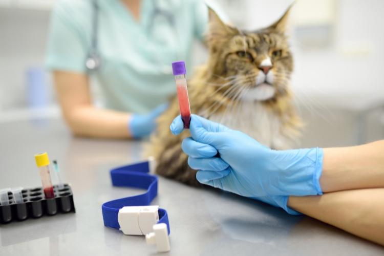
Diagnostic imaging refers to non-invasive techniques that create visual representations of the inside of your pet’s body. These images help veterinarians examine organs, bones, tissues, and internal structures without surgery.
During an imaging procedure, your veterinarian gathers detailed information that may not be visible during a standard wellness exam. Because many conditions develop internally, imaging often provides the clarity needed for early and accurate diagnosis.
Why Diagnostic Imaging Is So Important
Although physical exams reveal valuable information, they have limitations. Some conditions, such as internal injuries or organ abnormalities, cannot be detected through touch or observation alone. Therefore, diagnostic imaging fills this gap.
More importantly, imaging allows veterinarians to:
- Detect hidden injuries or abnormalities
- Confirm or rule out suspected conditions
- Monitor disease progression
- Evaluate treatment effectiveness
- Plan surgical procedures with precision
Because imaging improves diagnostic accuracy, it reduces guesswork and leads to more targeted care.
Common Types of Diagnostic Imaging for Pets
Veterinary medicine uses several imaging techniques, each serving a unique purpose. Together, they provide a comprehensive view of your pet’s health.
X-Rays (Radiographs)
X-rays are one of the most widely used imaging tools in veterinary care. They create images of dense structures, such as bones and certain organs.
Veterinarians commonly use X-rays to diagnose:
- Bone fractures and joint problems
- Arthritis and spinal issues
- Heart and lung conditions
- Foreign objects in the digestive tract
Because X-rays are quick and effective, they often serve as the first step in diagnosing many health concerns.
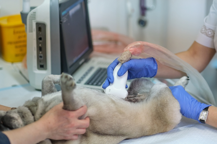
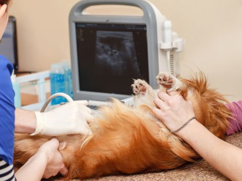
Ultrasound
Unlike X-rays, ultrasound uses sound waves to produce real-time images of soft tissues and organs. This technique allows veterinarians to observe movement and blood flow within the body.
Ultrasound is especially useful for examining:
- Abdominal organs, such as the liver and kidneys
- The heart and circulatory system
- Pregnancy and fetal development
- Fluid accumulation or internal masses
Additionally, ultrasound often helps guide minimally invasive procedures, such as biopsies.
Advanced Imaging Techniques
In certain cases, veterinarians may recommend advanced imaging, such as CT scans or MRI. These tools provide highly detailed, cross-sectional images of the body.
Advanced imaging is particularly valuable for diagnosing:
- Neurological conditions
- Complex joint injuries
- Tumors and internal growths
- Chronic or unexplained pain
Although these techniques are more specialized, they significantly enhance diagnostic precision when standard imaging is not sufficient.
Early Detection Leads to Better Outcomes
One of the greatest advantages of diagnostic imaging is early detection. Many serious conditions, including cancer, heart disease, and organ dysfunction, begin with subtle changes that are invisible externally.
However, imaging allows veterinarians to identify these changes early. As a result, treatment can begin sooner, which often improves recovery and long-term prognosis.
For example, detecting a small mass before it grows larger may allow for less invasive treatment. Similarly, identifying joint degeneration early helps manage pain and mobility before irreversible damage occurs.
Diagnostic Imaging Supports Accurate Treatment Planning
Accurate diagnosis is the foundation of effective treatment. Without imaging, veterinarians may need to rely on trial-and-error approaches. Imaging eliminates uncertainty by providing clear visual evidence of the problem.
With this information, veterinarians can:
- Choose the most appropriate treatment options
- Avoid unnecessary procedures
- Monitor healing and response to therapy
- Adjust treatment plans as needed
Therefore, diagnostic imaging not only identifies problems but also ensures that pets receive the right care at the right time.
Safe and Minimally Invasive Procedures
Many pet owners worry about the safety of diagnostic imaging. Fortunately, most imaging techniques are non-invasive and safe when performed by trained professionals.
For instance, X-rays use minimal radiation, and veterinarians take precautions to protect both pets and staff. Ultrasound involves no radiation at all and causes little to no discomfort. In some cases, mild sedation may be used to keep pets calm and still, ensuring accurate images.
Overall, the benefits of diagnostic imaging far outweigh the risks, especially when it leads to early and effective treatment.
Imaging Helps Monitor Chronic Conditions
For pets with chronic illnesses, diagnostic imaging plays an ongoing role in care. Conditions such as arthritis, heart disease, or kidney problems often require regular monitoring.
Through repeat imaging, veterinarians can track disease progression and evaluate how well treatments are working. This proactive approach helps maintain your pet’s comfort and quality of life over time.
When Might Your Pet Need Diagnostic Imaging?
Your veterinarian may recommend diagnostic imaging if your pet shows signs such as:
- Persistent limping or pain
- Unexplained weight loss
- Difficulty breathing
- Vomiting or diarrhea that does not resolve
- Behavioral changes or lethargy
Even when symptoms seem mild, imaging can uncover underlying issues that require attention.
Diagnostic imaging is a powerful tool that allows veterinarians to see what cannot be detected during a physical exam alone. By providing clear, detailed views of internal structures, imaging supports early detection, accurate diagnosis, and effective treatment planning. Ultimately, it improves outcomes and helps pets live healthier, more comfortable lives.
If you are seeking advanced and reliable pet diagnostic care in South Burlington, VT, diagnostic imaging is an essential part of ensuring your pet receives timely, precise, and compassionate veterinary care.
Frequently Asked Questions
Q1. Is diagnostic imaging safe for pets?
Ans. Yes. Most imaging procedures are non-invasive and safe. Veterinarians follow strict safety protocols to minimize risks.
Q2. Do pets need sedation for imaging?
Ans. Sometimes. Mild sedation may be used if a pet is anxious or unable to remain still, but many imaging procedures do not require it.
Q3. How long does diagnostic imaging take?
Ans. X-rays typically take only a few minutes, while an ultrasound may take slightly longer, depending on the area being examined.
Q4. Can diagnostic imaging detect cancer in pets?
Ans. Imaging can identify masses or abnormalities that may indicate cancer. Additional tests are often needed for confirmation.
Q5. How often should pets undergo diagnostic imaging?
Ans. Imaging is recommended as needed based on symptoms, age, and health conditions. Senior pets may require more frequent imaging.
Q6. Does diagnostic imaging replace physical exams?
Ans. No. Imaging complements physical exams by providing internal insights that support a complete health evaluation.